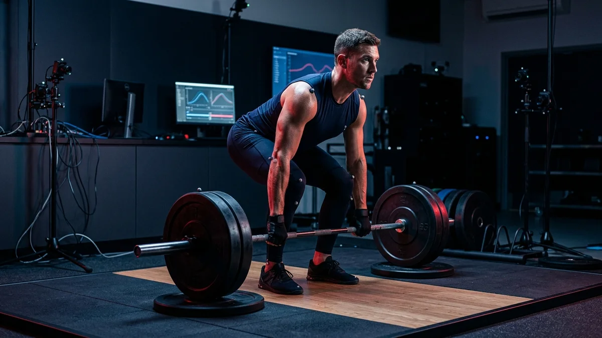
1. Anatomy & Biomechanical Definition of the Hip Hinge
The hip hinge is a fundamental multi-joint pattern characterized by posterior pelvic translation and maximal flexion at the acetabulofemoral joints with minimal knee bend (15 to 20 degrees). This posture protects the lumbar spine from harmful shear forces while maximally loading the gluteus maximus, hamstrings, and erector spinae.
Unlike a standard knee-dominant bilateral squat—where the femur and tibia both rotate through substantial angular displacement and the center of mass drops primarily downward—the hip hinge is purely hip-dominant. The tibia remains virtually perpendicular to the platform, shifting the ground reaction force vector directly behind the midfoot and loading the powerful posterior kinetic chain.
At the anatomical center of this movement is the acetabulofemoral ball-and-socket joint. When executed properly, the pelvis rotates forward over the femoral heads in an anterior-posterior arc. Because the human pelvis and sacrum form the mechanical foundation for the lumbar vertebrae (L1 through L5), stabilizing the lumbopelvic complex during this rotation is what distinguishes an elite, injury-resilient deadlift from a catastrophic lumbar disc herniation.
2. Sagittal Plane Moment Arms & Load Distribution
Every strength exercise is governed by rotational torque (τ = F × r), where F represents the gravitational resistance of the barbell plus upper body mass, and r represents the perpendicular moment arm length from the axis of rotation to the line of gravitational action.
In the sagittal plane of a deadlift or Romanian deadlift (RDL), there are three primary anatomical rotational fulcrums:
- The Lumbosacral Fulcrum (L5/S1): The perpendicular distance from the barbell to the lumbosacral junction creates an immense extensor torque demand on the lumbar erector spinae (longissimus thoracis, iliocostalis lumborum, and multifidus).
- The Acetabulofemoral Fulcrum (Hips): The horizontal distance from the barbell to the hip joint centers creates the primary hip extensor moment, loaded onto the gluteus maximus and adductor magnus.
- The Tibiofemoral Fulcrum (Knees): In a pure hinge, knee flexion is restricted to approximately 15° to 20°, keeping the quadriceps moment arm minimal compared to a squat.
Crucially, if the barbell drifts even 3 centimeters anteriorly away from the shins during the pull, the moment arm to the lumbosacral junction (L5/S1) increases exponentially. This introduces hundreds of additional Newton-meters of flexor torque onto the lumbar spine, forcing the erector spinae into mechanical disadvantage.
To counteract this, lifters must recruit the latissimus dorsi. By isometric adduction and internal rotation of the humerus ("tucking the armpits into the back pockets"), the lats actively pull the barbell posterior toward the tibia, shortening the external lumbar moment arm by 3 to 5 cm and slashing lumbar spinal stress by up to 25%.
3. Lumbar Shear Force vs Compressive Load: The McGill Matrix
Dr. Stuart McGill, professor emeritus of spine biomechanics at the University of Waterloo, conducted seminal cadaveric and in vivo research evaluating human spinal tissue tolerance under load. His laboratory established a vital distinction that every lifter must memorize:
The human lumbar spine in neutral lordosis can easily tolerate over 12,000 Newtons (~2,700 lbs) of pure axial compressive load without structural failure. However, when the lumbar spine flexes under load, its tolerance to anterior-posterior shear force drops precipitously to under 2,000 Newtons (~450 lbs).
When you maintain a neutral spine during a heavy deadlift (approaching your 1RM), the load is transferred almost entirely as axial compression along the thick vertebral bodies and hydrated cartilaginous intervertebral discs. The spinal erector muscle fascicles attach obliquely to the vertebrae at angles that create counteracting posterior shear forces, neutralizing the anterior shear induced by the forward-leaning torso.
However, once the lumbar spine enters flexion (rounding the lower back):
- The muscle fascicles of the erector spinae re-orient parallel to the spine, completely losing their ability to counteract anterior shear.
- The active musculature experiences electromyographic silence (the "flexion-relaxation phenomenon"), offloading the load onto passive viscoelastic structures: the supraspinous ligament, interspinous ligament, and posterior fibers of the annulus fibrosus.
- Hydrostatic pressure within the nucleus pulposus surges backward against the thinned posterior annular wall, leading to hydraulic extrusion, micro-tearing, and acute disc herniation.
To safeguard the spine, lifters must generate Intra-Abdominal Pressure (IAP) via the Valsalva maneuver. Contracting the diaphragm downward while bracing the transverse abdominis, internal obliques, and pelvic floor expands the abdominal cavity outward against a rigid lifting belt, creating a hydraulic cylinder that reduces axial spinal compression by 20% to 40%.
4. Pelvic Tilt, Hamstring Tension & Acetabulofemoral Geometry
The hamstrings (biceps femoris long head, semitendinosus, and semimembranosus) are bi-articular muscles originating at the ischial tuberosity of the pelvis and inserting below the knee. Because they cross both the hip and knee joints, their length-tension relationship is intimately coupled to pelvic orientation.
During the eccentric descent of a hip hinge, the pelvis must anteriorly rotate relative to the femurs (anterior pelvic tilt in space) while the lumbar spine remains locked in neutral. This dynamically stretches the hamstring complex at its proximal origin, storing elastic strain energy in the collagenous tendons and titin proteins of the muscle sarcomeres.
If an athlete possesses restricted hamstring extensibility or lacks kinesthetic awareness, their hamstrings reach passive insufficiency before full hinge depth is reached. When the hamstrings run out of length, they act as a rigid tether, pulling the ischial tuberosity forward and forcing the pelvis into posterior pelvic tilt ("butt wink"). The moment the pelvis tilts posteriorly, the lumbar spine is forcefully dragged into flexion, exposing the L4-L5 and L5-S1 motion segments to dangerous shear force.
5. The 4-Step Somatic Progression: Wall Tap to Barbell RDL
Mastering the hip hinge requires reprogramming motor unit firing patterns in the cerebellum. The following four-stage clinical progression takes lifters from sensory awareness to heavy loaded execution:
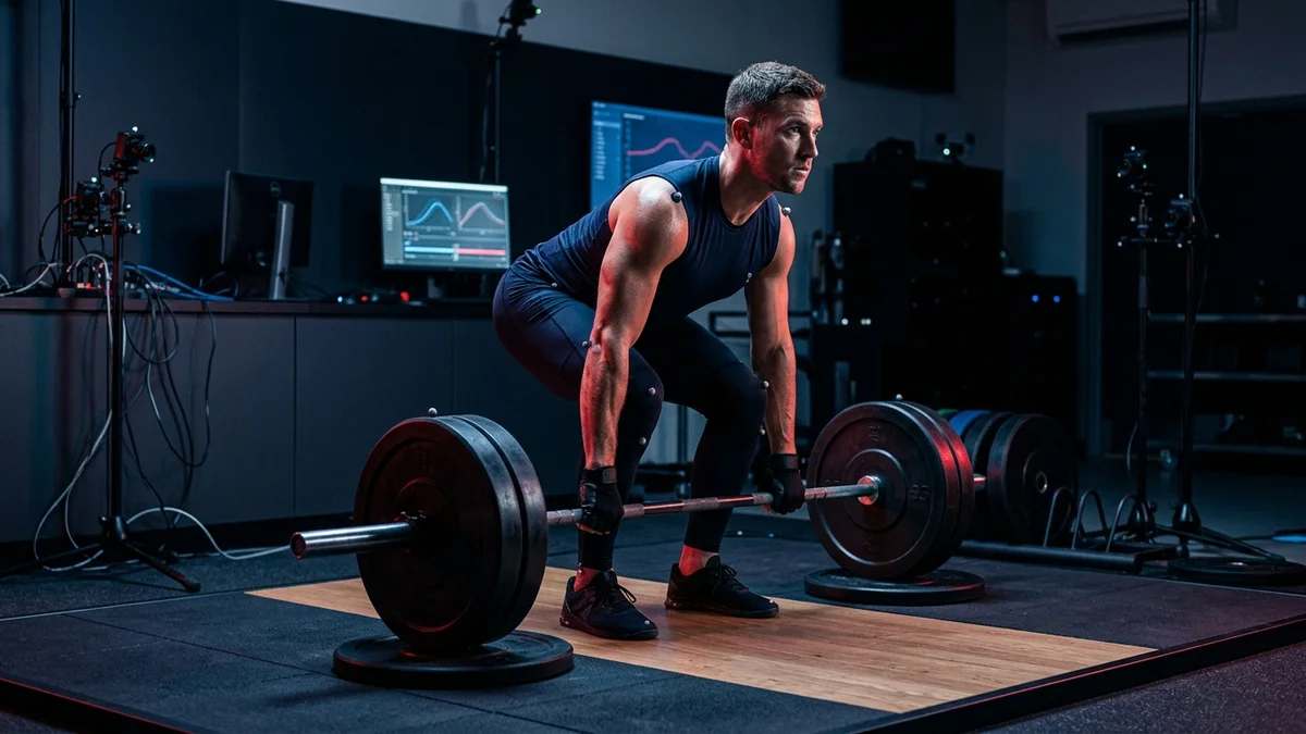
Drill 1: The 3-Point Dowel Rod Tactile Drill
Setup: Hold a light wooden dowel along your spine vertically. One hand rests in the cervical lordotic curve; the other rests in the lumbar lordotic curve.
Execution: The dowel must remain in constant contact with three anatomical landmarks at all times: (1) the sacrum, (2) the thoracic spine (between shoulder blades), and (3) the posterior occiput of the head. Unlock your knees 15° and push your hips backward. If the dowel loses contact with your sacrum, your lumbar spine has rounded; if it leaves your head, you are hyperextending your neck.
Drill 2: The Wall-Tap Hip Displacement Drill
Setup: Stand facing away from a wall, roughly 6 inches (15 cm) in front of baseboard. Feet shoulder-width apart, toes pointing forward or slightly flared (5-10°).
Execution: Place your hands in the crease of your hips. Maintain vertical shins and push your buttocks straight backward until your glutes touch the wall. Do not drop your hips downward; think about closing an open car door with your hips. Once mastered, step forward another 2 inches and repeat.
Drill 3: The Kettlebell Deadstop Hinge
Setup: Place a moderate kettlebell (16 to 24 kg) directly between the arches of your feet, aligned with the medial malleoli.
Execution: Hinge backward until your hands grasp the kettlebell handle. Engage your lats by squeezing your armpits, fill your abdomen with 360-degree air, and drive your hips forward into the bell to stand tall. Return the kettlebell to a complete dead stop between your feet on every repetition.
Drill 4: The Barbell Romanian Deadlift (RDL)
Setup: Unrack a loaded barbell (calculate plates with our Barbell Plate Loader) from standing lockout. Establish a solid grip just outside your thighs.
Execution: Inhale, brace IAP, unlock knees slightly, and trace the barbell along your thighs and over your patellae down toward the mid-shin. Stop the descent the instant your hips cease moving backward. Drive through the midfoot and snap your glutes to full lockout without hyperextending your lumbar spine.
6. Common Biomechanical Faults & Real-Time Corrections
Even seasoned strength athletes frequently commit subtle kinematic errors that compromise mechanical efficiency. Here is how to diagnose and correct the five most prevalent hip hinge breakdowns:
| Kinematic Fault | Biomechanical Mechanism | Injury Risk | Evidence-Based Cue & Correction |
|---|---|---|---|
| Squatting the Hinge | Excessive forward tibial translation (>30° knee angle) shifting load to patellar tendon and quads. | Sub-optimal posterior chain stimulus; patellofemoral shear. | "Lock shins vertical like tree trunks; push your hips back toward the far wall." |
| Lumbar Flexion (Rounding) | Erector spinae failure, passive ligamentous loading, and posterior disc bulging. | High risk of acute L4/L5 or L5/S1 disc herniation and annular tears. | "Show the room your chest logo; pack lats like squeezing oranges in your armpits." |
| Lockout Hyperextension | Leaning upper torso backward at top of lift using lumbar spine rather than glute hip extension. | Facet joint impingement, spondylolysis, and pars interarticularis stress fractures. | "Stand tall like a soldier; finish with glute squeeze, ribs down, pelvis neutral." |
| Cervical Hyperextension | Craning the neck upward to stare at the gym mirror during bottom of deadlift/RDL. | Cervical facet compression, trapezius strain, and vestibular disorientation. | "Pack a double chin; keep gaze focused on floor 8-10 feet in front of your toes." |
| Bar Drifting Anteriorly | Lats unengaged; barbell drifts 5-10 cm away from legs, massively increasing L5/S1 moment arm. | Exponential surge in spinal shear force; blown lockout mechanics. | "Paint your shins and thighs with the bar; bend the barbell around your legs." |
7. Comparative Biomechanical Loading Matrix
Different hip hinge and pulling variations distribute rotational torque and shear force across distinct segments of the musculoskeletal framework. The matrix below outlines how key exercises compare:
| Exercise Variation | Hip Moment Arm | Knee Moment Arm | Spinal Shear Stress | Primary Kinetic Target |
|---|---|---|---|---|
| Conventional Deadlift | High (25–30 cm) | Moderate (10–15 cm) | Moderate–High | Gluteus Maximus, Hamstrings, Erector Spinae, Quads |
| Romanian Deadlift (RDL) | Very High (30–35 cm) | Minimal (5–8 cm) | Moderate | Semitendinosus, Biceps Femoris, Gluteus Medius/Maximus |
| Trap Bar Deadlift (High Handle) | Moderate (18–22 cm) | High (15–20 cm) | Lowest (Aligned with CoM) | Vastus Lateralis/Medialis, Gluteus Maximus, Trapezius |
| Low-Bar Back Squat | Moderate–High (20–25 cm) | High (18–22 cm) | Moderate | Quadriceps, Gluteus Maximus, Adductor Magnus |
8. Peer-Reviewed Citations
- McGill, S. M. (2002). Low back disorders: Evidence-based prevention and rehabilitation. Human Kinetics Publishers, Champaign, IL.
- Cholewicki, J., & McGill, S. M. (1996). Mechanical stability of the in vivo lumbar spine: implications for injury and chronic lack of stability. Clinical Biomechanics, 11(1), 1–15.
- Escamilla, R. F., Francisco, A. C., Fleisig, G. S., et al. (2000). A three-dimensional biomechanical analysis of sumo and conventional style deadlifts. Medicine & Science in Sports & Exercise, 32(7), 1265–1275.
- Vigotsky, A. D., Harper, E. N., Ryan, D. R., et al. (2017). Biomechanical Comparison of the Hexagonal Barbell and Straight Barbell Deadlift. Journal of Strength and Conditioning Research, 31(3), 562–574.
- Swinton, P. A., Stewart, A., Agouris, I., et al. (2011). A biomechanical analysis of straight and hexagonal barbells using submaximal loads. Journal of Strength and Conditioning Research, 25(7), 2000–2009.
- Schoenfeld, B. J., Contreras, B., Tiryaki-Sonmez, G., et al. (2014). Regional Differences in Muscle Activation During Hamstrings Exercise. Journal of Strength and Conditioning Research, 29(1), 159–164.
9. Frequently Asked Questions
Why does my lower back get sore after Romanian deadlifts instead of my hamstrings?
Lower back soreness after RDLs usually occurs because you are hinging past your active hamstring flexibility threshold. Once your hamstrings reach full stretch, continuing downward forces your pelvis to tilt posteriorly and your lumbar spine to round, placing massive shear load on the lumbar erectors.
Is a rounded upper back (thoracic flexion) acceptable during maximum deadlifts?
Controlled thoracic flexion is common among competitive powerlifters because it shortens the moment arm between the barbell and the hips. However, thoracic flexion is only acceptable if the lumbar spine remains rigidly locked in neutral lordosis; lumbar flexion under maximal loads dramatically increases disc herniation risks.
How do I know if I have adequate hamstring mobility for a conventional deadlift from the floor?
Perform an active straight leg raise lying supine. If you cannot raise one leg to 75–80 degrees while keeping the opposite leg flat on the floor and your lumbar spine in neutral, pulling from the floor will likely force lumbar flexion. Elevate the bar on 2-4 inch blocks until mobility improves.
What tools on FitNet USA can help me calculate loads for hip hinge exercises?
Use our interactive One-Rep Max Calculator to compute your percentage training zones (70%–85% 1RM) and our Barbell Plate Loader to determine the exact plate configuration on Olympic barbells.
Should I wear a weightlifting belt during hip hinge drills?
During light motor-learning drills (dowel rod, wall taps, sub-60% loads), train without a belt to build intrinsic neuromuscular recruitment of the transversus abdominis and obliques. When working with loads exceeding 80% 1RM or performing heavy sets near failure, a 10mm or 13mm leather belt significantly increases intra-abdominal pressure and spinal stability.